An Ophthalmoscope Is Used to Determine Which of the Following
This should be followed by a distant direct examination at 22-25cm a comfortable near vision distance. Red reflex anterior segment disc vessels and lastly macula see box.

Moran Core How To Use The Direct Ophthalmoscope
HOME MEDICAL STUDENT USE.
. The first step in the use of an ophthalmoscope is to do examination at 1m distance. In a typical attack a small central disturbance in the field of vision marches toward the periphery leaving a transient scotoma in its wake. Have a longer useful life d.
Which aperture would be used to assess the eyes of a patient with undilated pupils C small at the conclusion of the examination the examiner shouldD summarize findings to the patient When the practitioner enters the examining room the infant patient is asleep. Direct ophthalmoscopy the most common procedure involves the use of a handheld tool called an ophthalmoscope to look inside the eye. Provide more accurate representation of color A.
It is used to detect and evaluate symptoms of various retinal vascular diseases or eye diseases such as glaucoma. Ophthalmoscope pictures fundus photographs and ultrasonographic findings supplemented by a fundus diagram provide the foundation for determining tumor size verifying tumor location and surrounding critical structures. You will be seated in a darkened room.
What am I looking for. Assessing skin with the use of pads of fingertips. Detect foreign bodies in the cornea b.
Placing the patient in a supine position. Use an ophthalmoscope to watch the clients pupil constrict when a strong light is shown on it. 447 This usually occurs with a visual aura lasting about 20 min.
The ophthalmoscope can be used to. Washing hands under warm water. An ophthalmoscope is an instrument that has a light and several small lenses on it.
Most ophthalmologists have deserted the direct ophthalmoscope for a different device an indirect ophthalmoscope which provides a better view of the peripheral retina and assists in the discovery of retinal holes etc. The health care provider performs this exam by shining a beam of light through the pupil using an instrument called an ophthalmoscope. The large light is best if.
You will either lie. They may ask you to look in certain directions. Ophthalmoscopes usually have 2 or 3 sizes of light to use depending on the level of pupil dilation.
An expected part of every eye exam ophthalmoscopes are. Well lit room no pupil dilators used. This tool has special lenses and a bright light that enable your doctor to focus past your pupil and onto the back of your eye.
A preliminary evaluation of adjacent critical structures can. Perfect for DIY At-Home ENT Doctor Visit or for nurse practitioner other medical students. An ophthalmoscope is particularly useful for examining the structures of the retinathe light sensitive area at the back of the eye responsible for processing images.
The diagnosis is confirmed by ophthalmoscopic examination of the dilated eye. Have higher light intensity b. Classic Migraine See also Chap.
Traditionally part of almost every eye exam ophthalmoscopes can identify healthy structures within the eyeball and easily help your eye doctor see symptoms or indicators of diseases of the eye. They are used all over the world and are an essential piece of apparatus for all who wish to study the intricate biology of the eye. The ophthalmoscope can also be used for examining the anterior part of the eye by turning the lens dial to 10.
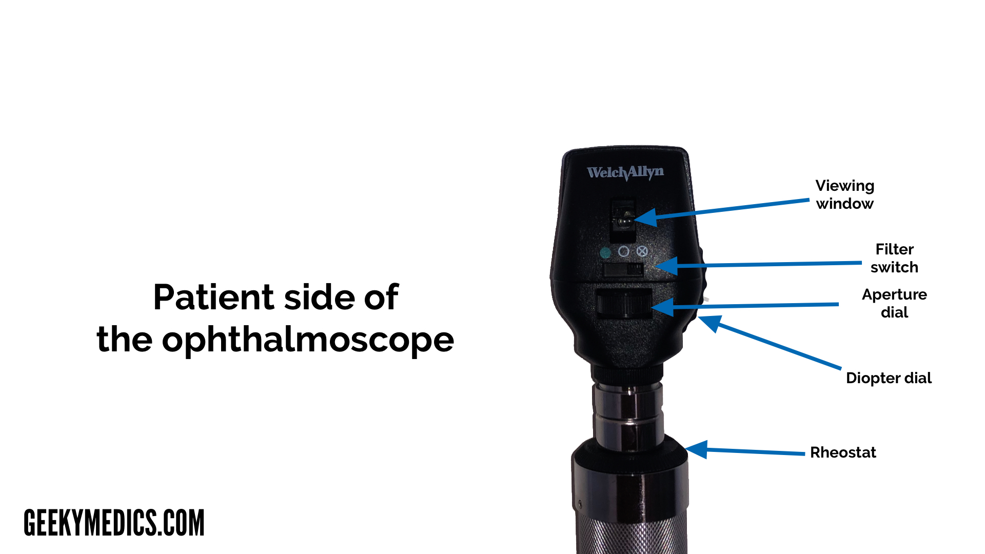
447 This usually occurs with a visual aura lasting about 20 min. The ophthalmoscope has 5 apertures. It has a light and different tiny lenses that allow the provider to view the back of the eyeball.
This sheds light on any abnormalities of the eyelids orbit and periorbita as well as highlights any obvious ocular deviations. The diagnosis is confirmed by ophthalmoscopic examination of the dilated eye. List of 10 Best ophthalmoscope.
In a typical attack a small central disturbance in the field of vision marches toward the periphery leaving a transient scotoma in its wake. All of the above B Halogen illumination does not a. ENT Diagnostic Kit 325V LED Otoscope Ophthalmoscope ENT Diagnostic Kit.
Detect lens opacities d. The practitioner would best. Your eye doctor can look through the lenses to examine your eye.
The ophthalmoscope has 5 apertures which aperture. An ophthalmoscope is about the size of a flashlight. Darken with use c.
Ophthalmoscopy is done as part of a routine physical or complete eye examination mainly done by optometrists and ophthalmologists. Verify doubtful papillary action c. An ophthalmoscope is a piece of equipment utilised by ophthalmologists that are used to inspect the internal structure of your eyes containing the retina.
Ophthalmoscopy also called fundoscopy is an exam your doctor optometrist or ophthalmologist uses to look into the back of your eye. Arrange the steps of palpation in the order the nurse would perform them during a patients physical examination. An ophthalmoscope can be used to check for.
With it they can see the retina which senses light and. In an exam once you have found an abnormality keep looking for a second one. The small light is used when the pupil is very constricted ie.
Damage to the optic nerve Retinal tear or detachment Glaucoma Macular degenerations Melanoma Diabetic retinopathy Hypertension Infection More advanced ophthalmoscopes offer doctors the ability to alter the aperture lens and aperturefilter combinations to gain a larger view of the fundus. Classic Migraine See also Chap. Sit facing the client and while look directly at the client.
Fundoscopy Ophthalmoscopy Osce Guide Geeky Medics
Fundoscopy Ophthalmoscopy Osce Guide Geeky Medics
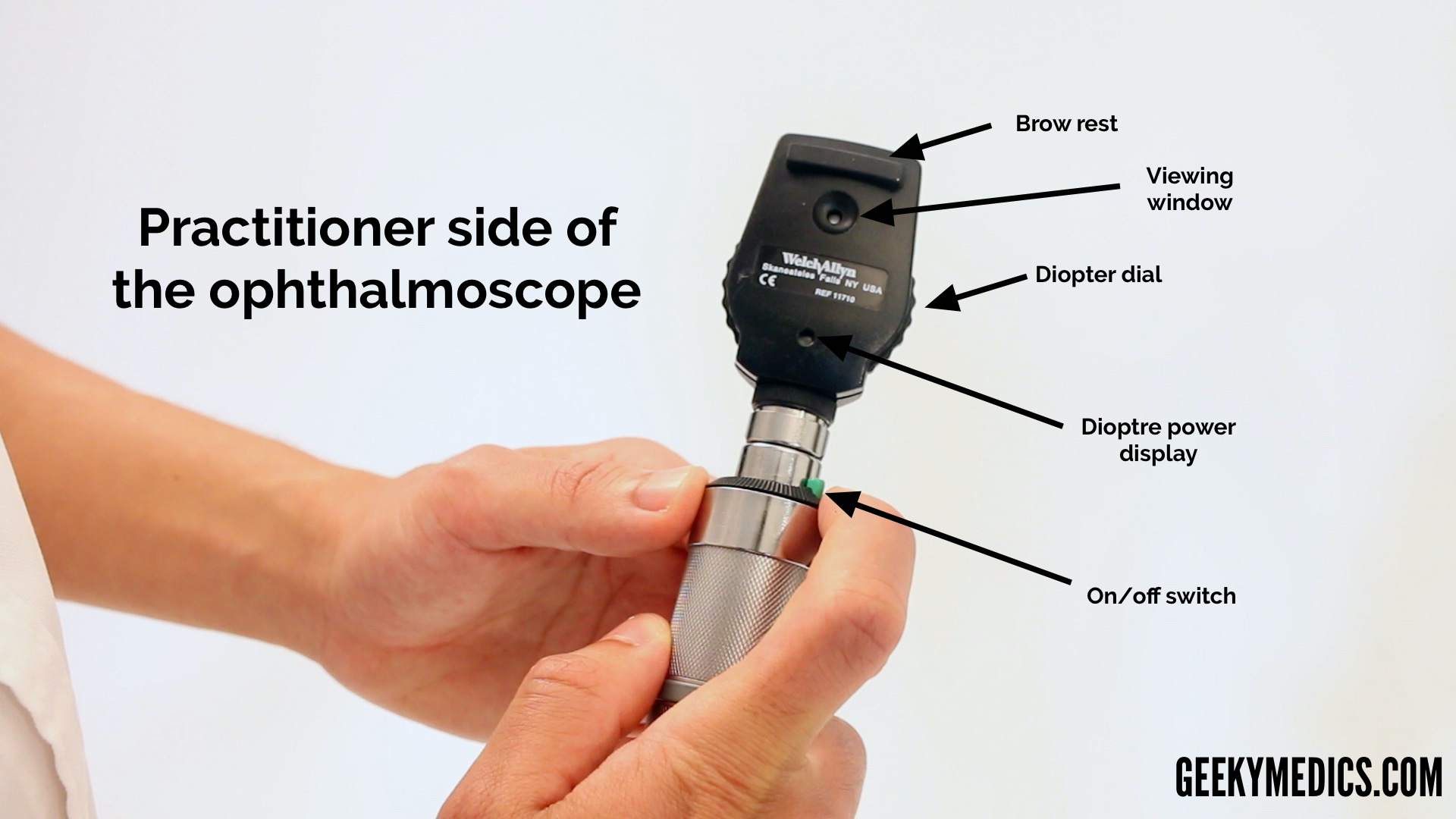
Fundoscopy Ophthalmoscopy Osce Guide Geeky Medics
No comments for "An Ophthalmoscope Is Used to Determine Which of the Following"
Post a Comment